Therapy For Bursitis Of The Foot
Overview
Retrocalcaneal bursitis is closely related to Haglund?s Deformity (or ?pump bumps?). If you have a bony enlargement on the back of the heel that rubs the Achilles tendon, it can cause the formation of a bursa (small fluid filled sack). It usually happens in athletes as shoes rub against the heel. The bursa can aggravated by the stitching of a heel counter in the shoe as well. It can make wearing shoes and exercising difficult. Another term used for this condition is ?pump bump? because it can frequently occur with wearing high heels as well. ?Retro-" means behind and ?calcaneus? means heel bone. So this is precisely where the bursitis (inflammation of the bursa) develops. Once it begins and you develop bursitis between the heel bone and the Achilles tendon, it can become even more painful. When most people first notice retrocalcaneal bursitis, it is because the skin, bursa and other soft tissues at the back of the heel gets irritated as the knot of bone rubs against the heel counter in shoes. The back of the shoes create friction and pressure that aggravate the bony enlargement and pinches the bursa while you walk.
Causes
Bursitis can be caused by an injury, an infection, or a pre-existing condition in which crystals can form in the bursa. Injury. An injury can irritate the tissue inside the bursa and cause inflammation. Doctors say that bursitis caused by an injury usually takes time to develop. The joints, tendons, or muscles that are near the bursae may have been overused. Most commonly, injury is caused by repetitive movements.
Symptoms
Medical experts strongly recommend that you consult a doctor if you have any of the symptoms below. Disabling joint pain that prevents you from doing your daily activities. Pain that lasts for more than two weeks. Excessive swelling, redness, bruising or a rash around the painful joint. Sharp or shooting pain, especially when you exercise or do something more strenuous. A fever. Any of the above could be a sign of infection, a condition such as arthritis or a more serious injury such as a tendon tear that may require medical attention.
Diagnosis
After you have described your foot symptoms, your doctor will want to know more details about your pain, your medical history and lifestyle, including whether your pain is worse at specific times of the day or after specific activities. Any recent injury to the area. Your medical and orthopedic history, especially any history of diabetes, arthritis or injury to your foot or leg. Your age and occupation. Your recreational activities, including sports and exercise programs. The type of shoes you usually wear, how well they fit, and how frequently you buy a new pair.
Non Surgical Treatment
Many times, Achilles tendon bursitis can be treated with home care. However, if self-care remedies do not work, your doctor may need to administer additional treatments. Home care. The most important thing you can do to help your healing is to refrain from activities that could further aggravate the bursitis. Typical home-care treatments for Achilles tendon bursitis include Anti-inflammatory medication. Take over-the-counter pain medication, like aspirin or ibuprofen, to reduce pain and swelling. Heel wedges. An over-the-counter or custom heel wedge can be placed in your shoe to minimize stress in the Achilles tendon. Ice. Apply ice or other cold therapy to your sore heel several times a day. Rest. Limit your activity on the injured leg, avoid putting pressure on it whenever possible. Also, elevate your leg during non-use to help reduce the swelling. The above remedies may be used on their own or in combination with others on the list. Physician-administered treatments. If the above self-care remedies are not effective, you should visit your doctor for additional treatment. Possible Achilles tendon bursitis treatments your doctor may administer include immobilization. If the bursitis is combined with Achilles tendonitis, your doctor may place a temporary cast on the ankle to prevent movement and allow it to heal. Physical therapy. Exercises may be used to improve the ankle's strength and flexibility. Steroids. Injection of steroids into the retrocalcaneal bursa (not the Achilles tendon) may be necessary. Surgery. In very rare circumstances, surgery may be needed to remove the retrocalcaneal bursa, however, this is typically a last resort.
Surgical Treatment
Surgery is rarely need to treat most of these conditions. A patient with a soft tissue rheumatic syndrome may need surgery, however, if problems persist and other treatment methods do not help symptoms.
Prevention
Maintain proper form when exercising, as well as good flexibility and strength around the ankle to help prevent this condition. Proper stretching of the Achilles tendon helps prevent injury.
Rigid Hammertoes Treatments
 Overview
The term, Hammer toe, is commonly used as a general classification for any condition where the toe muscle weakens, causing digital contracture, and resulting hammertoes in deformity, a digital contracture like this can actually be a hammertoe, claw toe or mallet toe, depending on which joints in the toe are contracted. Clawtoes are bent at the middle and end joints, while hammertoes are bent at the middle joint only. When it?s mallet toe, the joint at the end of the toe buckles. The skin near the toenail tip develops a painful corn that can eventually result in an ulcer. Doctors further categorize all forms of hammertoe based on whether the affected toe is flexible, semi-rigid or rigid. The more rigid the toe, the more pain it will cause.
Causes
Some causes of hammertoe are shoes that are too tight or short, shoes with high heels, injury, Diseases that affect the nerves and muscles, such as arthritis and diabetes. When shoes do not fit well, over time the pressure of the shoes pushes the toes into a bent position. After a while, the muscles become unable to straighten the toe, even when you are not wearing shoes. Similarly, when there is damage or disease of the nerves or muscles in the toes, the toe may rest in the bent position until the tendons become permanently shortened and the toe becomes a rigid hammertoe. The risk of developing a hammertoe increases with age. Women are much more likely to develop a hammertoe than men.
 Symptoms
Signs and symptoms of hammertoe and mallet toe may include a hammer-like or claw-like appearance of a toe. In mallet toe, a deformity at the end of the toe, giving the toe a mallet-like appearance. Pain and difficulty moving the toe. Corns and calluses resulting from the toe rubbing against the inside of your footwear. Both hammertoe and mallet toe can cause pain with walking and other foot movements.
Diagnosis
Although hammertoes are readily apparent, to arrive at a diagnosis the foot and ankle surgeon will obtain a thorough history of your symptoms and examine your foot. During the physical examination, the doctor may attempt to reproduce your symptoms by manipulating your foot and will study the contractures of the toes. In addition, the foot and ankle surgeon may take x-rays to determine the degree of the deformities and assess any changes that may have occurred.
Non Surgical Treatment
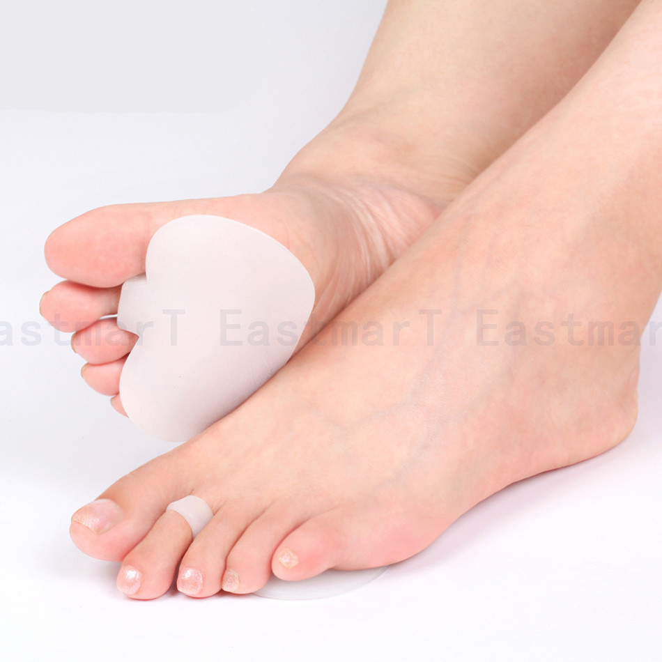
Wear wide shoes with plenty of room in the toes and resilient soles. Avoid wearing shoes with pointed toes. Commercially available felt pads or cushions may ease pressure from the shoe on the toe. Toe caps (small, padded sleeves that fit around the tip of the toe) may relieve the pain of hammer toe. Do toe exercises, to help toe muscles become stronger and more flexible.
Arch supports or an orthotic shoe insert prescribed by your doctor or podiatrist may help to redistribute weight on the foot. These devices do not cure the problem but may ease the symptoms of either hammer toe or mallet toe.
Surgical Treatment
If conservative measures fail to provide relief, or if your hammertoe is in advanced stages with rigidity and a significant amount of pain, surgery may be required. Some patients also require surgery if they have open sores or wounds related to their hammertoe. For patients who also suffer from bunions, a combined procedure may be appropriate, addressing both conditions within the same surgery. Recovery time will vary from patient to patient, depending on the extent of the surgical repair and other conditions that may also be present.
 Prevention
Some different things you can do to help prevent hammer toes from forming and progressing do exist. You can wear supportive shoes to help prevent deformities. Hammer toes are often times related to poor foot mechanics, particularly foot flattening. You can wear custom orthotics prescribed by a podiatrist; orthotics might prevent or slow the progression of hammer toes. You can also avoid shoes with narrow or pointed toe boxes that can compress your toes.
Physical Therapy After Bunion And Hammertoe Surgery
 Overview
Generally a hammertoe or mallet toe is caused by wearing high heels or shoes that are too small around the toe area, so it?s no surprise that it is mostly women who suffer from them. A Hammer toes has a bend in the middle joint of the toe whereas a mallet toe has a bend in the upper joint of the affected toe. The way someone walks (gait) can also lead to the formation of hammertoes and mallet toes as can overuse and injury. Sometimes a deep blister will form over the bent joint and often after some time calluses and corns will develop on the affected toe joint. People with arthritis, diabetes or neuromuscular conditions are also more likely to develop a hammer toe or mallet toe.
Causes
A hammertoe is formed due an abnormal balance of the muscles in the toes. This abnormal balance causes increased pressures on the tendons and joints of the toe, leading to its contracture. Heredity and trauma can also lead to the formation of a hammertoe. Arthritis is another factor, because the balance around the toe in people with arthritis is so disrupted that a hammertoe may develop. Wearing shoes that are too tight and cause the toes to squeeze can also be a cause for a hammertoe to form.
 Symptoms
For some people, a hammer toe is nothing more than an unsightly deformity that detracts from the appearance of the foot. However, discomfort may develop if a corn or callus develops on the end or top of the toe. If pressure and friction continue on the end or top of the toe, a painful ulcer may develop. Discomfort or pain can lead to difficulty walking.
Diagnosis
Although hammertoes are readily apparent, to arrive at a diagnosis the foot and ankle surgeon will obtain a thorough history of your symptoms and examine your foot. During the physical examination, the doctor may attempt to reproduce your symptoms by manipulating your foot and will study the contractures of the toes. In addition, the foot and ankle surgeon may take x-rays to determine the degree of the deformities and assess any changes that may have occurred.
Non Surgical Treatment
Your doctor will decide what type of hammertoe you have and rule out other medical conditions. Treatment may range from more appropriate footgear to periodic trimming and padding of the corn. Cortisone injections may be indicated if a bursitis is present. Antibiotics may be utilized in the presence of infection. Removable accommodative pads may be made for you.
Surgical Treatment
Toes can be surgically realigned and made straight again. They can even be made shorter. The good news is that toes can be corrected. Hammer toe surgery is often synonymous with ?toe shortening?, ?toe job? and/or ?toe augmentation?. Depending on the severity and length of the toe, there are several methods to surgically correct a hammer toe. In general, the surgery involves removing a portion of the bone Hammer toe at the contracted joint, to realign the toe.
 Prevention
Skin creams can help maintain skin softness and pliability. A pumice stone or loofah sponge can help get rid of dead skin. Taking a warm footbath for 10 minutes two or three times a week will keep the feet relaxed and help prevent mild foot pain caused by fatigue. Adding 1/2 cup of Epsom salts increases circulation and adds other benefits. Taking footbaths only when the feet are painful is not as helpful.
What Is A Tailor'S Bunion?
Overview
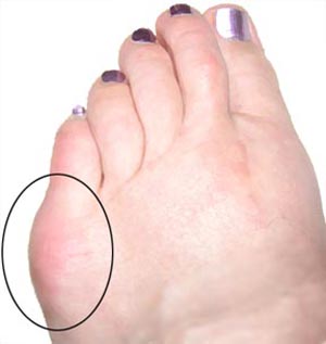 Bunion is the common term for a medical condition known as Hallux Valgus. Hallux Valgus is the tilting of the toe away from the mid-line of the body. It is usually characterized by a lump or bump that is red, swollen and/or painful on the inside of the foot in and around the big toe joint.
Causes
Bunions are most widely considered to be due to an imbalance in the forces that is exerted across the joint during walking. The resulting abnormal motion and pressure over the joint, over many years (combined with poor fitting footwear) leads to instability in the joint causing hallux valgus and bunions. Bunions are really only a symptom of faulty foot mechanics and are usually caused by a combination of the way we walk, the foot we inherit and inappropriate footwear use.
Symptoms
Look for an angular, bony bump on the side of the foot at the base of the big toe. Sometimes hardened skin or a callus covers this bump. There's often swelling, redness, unusual tenderness, or pain at the base of the big toe and in the ball of the foot. Eventually, the area becomes shiny and warm to the touch. Seek medical advice if you have persistent pain when walking normally in otherwise comfortable, flat-soled shoes, you may be developing a bunion, bursitis, or a bone spur in your foot.
Diagnosis
Before examining your foot, the doctor will ask you about the types of shoes you wear and how often you wear them. He or she also will ask if anyone else in your family has had bunions or if you have had any previous injury to the foot. In most cases, your doctor can diagnose a bunion just by examining your foot. During this exam, you will be asked to move your big toe up and down to see if you can move it as much as you should be able to. The doctor also will look for signs of redness and swelling and ask if the area is painful. Your doctor may want to order X-rays of the foot to check for other causes of pain, to determine whether there is significant arthritis and to see if the bones are aligned properly.
Non Surgical Treatment
Treatment may be surgical or non-surgical. The goal of non-surgical treatment is to relieve pressure on the foot and to prevent pressure sores and foot ulcers. This is accomplished by prescribing accommodative shoes with a wide toe box - sandals or extra depth shoes with soft moulded insoles. It may also be possible to relax the leather on shoes to make room for a bunion. 
Surgical Treatment
Sometimes a screw is placed in the foot to hold a bone in a corrected position, other times a pin, wire or plate is chosen. There are even absorbable pins and screws, which are used for some patients. In British Columbia, pins seem to be used most frequently, as they're easier to insert and less expensive. They are typically--but not always--removed at some point in the healing process. But as a general rule, Dr. Schumacher prefers to use screws whenever possible, as they offer some advantages over pins. First, using screws allows you to close over the wound completely, without leaving a pin sticking out of the foot. That allows for a lower infection rate, it allows you to get your foot wet more quickly following the surgery, and it usually allows for a quicker return to normal shoes. Second, they're more stable than pins and wires. Stability allows for faster, more uneventful, bone healing. Third, they usually don't need to be removed down the road, so there's one less procedure involved.
What Does Over-Pronation Mean
Overview
You know when we walk in the sand on the beach? Well, that?s how nature meant us to walk! You will notice how the sand forms a complete support under the foot. Unfortunately, instead of soft natural surfaces, we walk mainly on hard surfaces like pavements and floors. These hard surfaces cause the foot to roll inwards and the arches to flatten to gain ground contact. This condition is called ?over-pronation? and it affects an estimated 70% of the population. Over-pronation can lead to various foot problems such as heel pain and ball of foot pain. Because our feet are the foundation of our body, poorly aligned feet may also cause problems in other parts of the body, resulting in shin pain, aching legs, knee pain, even lower back pain.
Causes
There has been some speculation as to whether arch height has an effect on pronation. After conducting a study at the Rose-Hulman Institute of Technology, Maggie Boozer suggests that people with higher arches tend to pronate to a greater degree. However, the generally accepted view by professionals is that the most pronation is present in those with lower arch heights. To complicate matters, one study done by Hylton Menz at the University of Western Sydney-Macarthur suggests that the methods for measuring arch height and determining whether someone is ?flat-footed? or ?high-arched? are unreliable. He says, ?For this reason, studies investigating the relationship between static arch height motion of the rearfoot have consistently found that such a classification system is a poor predictor of dynamic rearfoot function.
Symptoms
Overpronation can lead to injuries and pain in the foot, ankle, knee, or hip. Overpronation puts extra stress on all the bones in the feet. The repeated stress on the knees, shins, thighs, and pelvis puts additional stress on the muscles, tendons, and ligaments of the lower leg. This can put the knee, hip, and back out of alignment, and it can become very painful.
Diagnosis
When you overpronate your foot rolls inwards causing the lower leg to rotate inwards too. It's thought that this increases stress on the soft tissues of the lower leg causing pain and inflammation, or more commonly known as shin splints.
Non Surgical Treatment
Side Step with Opposite Reach. This exercise is designed to load the "bungee cord system" of the gluteal muscle and its opposite, latissimus dorsi muscle to keep the foot from overpronating. Because the opposite arm swings across the front leg when walking, this exercise creates tension in the muscles all the way from the front foot, across the back of the hips and back, to the fingers of the opposite hand. Movement Directions. Stand with left foot on top of the dome of the BT. (Note: For added balance, the right foot can tap on the ground, if needed). Reach right leg out to the side of the BT, and tap the ground while squatting down on the left side and reaching right arm across the left knee. Push down with left big toe while squatting. This activates the arch of the left foot and strengthens all the stabilizing muscles on the left side of the lower body. Return to starting position. Perform 8 to 10 repetitions on each leg.
Prevention
Massage and stretch the calves to increase dorsiflexion at the foot/ankle. Dorsiflexion is the bending at the ankle. By improving the dorsiflexion, one will have more flexibility at the ankle, which will allow the foot to over-pronate less. Massage the IT Band with a foam roller or tennis ball to quiet down the tightness throughout this part of the leg. The IT Band attaches from the glute maximus and runs down the side of the leg into the knee area. When the IT Band is tight it will accelerate the force of the leg moving inward, which will cause the foot to move inward as well. It is often that tightness through the IT Band that promotes over-pronation. Decreasing over-pronation, which is very prominent in runners, will help add endurance, speed and efficiency to your run and ultimately place less stress on your body.
Rehab For Calcaneal Apophysitis
Overview
Sever?s disease is the most common cause of heel pain in children aged 9 to 14. Sever?s disease results from stress placed on the growth plate of the heel bone. An excessive amount of running or other activities can cause inflammation around the growth plate, which results in pain. Rest, ice and orthotics and proper shoes are usually prescribed.
Causes
Growth plates, also called epiphyseal plates, occur at the end of long bones in children who are still growing. These plates are at either end of growing bones, and are the place where cartilage turns into bone. As children grow, these plates eventually become bone (a process called ossification). During a growth spurt, the bone in the heel may outpace the growth of the muscles and tendons that are attached to the heel, such as the Achilles tendon. During weight bearing, the muscles and tendons begin to tighten, which in turn puts stress on the growth plate in the heel. The heel is not very flexible, and the constant pressure on it begins to cause the symptoms of Sever?s disease. Sever?s disease is common, and it does not predispose a child to develop any other diseases or conditions in the leg, foot, or heel. It typically resolves on its own.
Symptoms
Pain is usually felt at the back of the heel and around the sides of the heel. If you squeeze the back of the heel from both sides simultaneously and pain is experienced Sever?s disease is more than likely present.
Diagnosis
A doctor can usually tell that a child has Sever's disease based on the symptoms reported. To confirm the diagnosis, the doctor will probably examine the heels and ask about the child's activity level and participation in sports. The doctor might also use the squeeze test, squeezing the back part of the heel from both sides at the same time to see if doing so causes pain. The doctor might also ask the child to stand on tiptoes to see if that position causes pain. Although imaging tests such as X-rays generally are not that helpful in diagnosing Sever's disease, some doctors order them to rule out other problems, such as fractures. Sever's disease cannot be seen on an X-ray.
Non Surgical Treatment
Treatment may consist of one or more of the following. Elevating the heel. Stretching hamstring and calf muscles 2-3 times daily. Using R.I.C.E. (Rest, Ice, Compression, Elevation). Foot orthotics. Medication. Physical therapy. Icing daily (morning). Heating therapy. Open back shoe are best and avoid high heel shoe. The Strickland Protocol has shown a positive response in patients with a mean return to sport in less than 3 weeks. Further research into the anatomical and biomechanical responses of this protocol are currently being undertaken.
What Is Posterior Tibial Tendon Dysfunction
Overview
Adult Acquired Flatfoot (Posterior Tibial Tendon Dysfunction) is a painful, progressive deformity in adults. It results from a gradual stretch (attenuation) of the tibialis posterior tendon and the ligaments that support your foot?s arch. This stretching causes the tendon to lose strength and function. Many people have flat feet and do not experience pain. However, pain occurs with Adult Acquired Flatfoot because the tendons and ligaments have been torn. Once the vital ligaments and posterior tibial tendon are lost, there is no longer anything holding the arch of the foot in place. 
Causes
Damage to the posterior tendon from overuse is the most common cause for adult acquired flatfoot. Running, walking, hiking, and climbing stairs are activities that add stress to this tendon, and this overuse can lead to damage. Obesity, previous ankle surgery or trauma, diabetes (Charcot foot), and rheumatoid arthritis are other common risk factors.
Symptoms
Often, this condition is only present in one foot, but it can affect both. Adult acquired flatfoot symptoms vary, but can swelling of the foot's inner side and aching heel and arch pain. Some patients experience no pain, but others may experience severe pain. Symptoms may increase during long periods of standing, resulting in fatigue. Symptoms may change over time as the condition worsens. The pain may move to the foot's outer side, and some patients may develop arthritis in the ankle and foot.
Diagnosis
In diagnosing flatfoot, the foot & Ankle surgeon examines the foot and observes how it looks when you stand and sit. Weight bearing x-rays are used to determine the severity of the disorder. Advanced imaging, such as magnetic resonance imaging (MRI) and computed tomography (CAT or CT) scans may be used to assess different ligaments, tendons and joint/cartilage damage. The foot & Ankle Institute has three extremity MRI?s on site at our Des Plaines, Highland Park, and Lincoln Park locations. These extremity MRI?s only take about 30 minutes for the study and only requires the patient put their foot into a painless machine avoiding the uncomfortable Claustrophobia that some MRI devices create.
Non surgical Treatment
Because of the progressive nature of PTTD, early treatment is advised. If treated early enough, your symptoms may resolve without the need for surgery and progression of your condition can be arrested. In contrast, untreated PTTD could leave you with an extremely flat foot, painful arthritis in the foot and ankle, and increasing limitations on walking, running, or other activities. In many cases of PTTD, treatment can begin with non-surgical approaches that may include. Orthotic devices or bracing. To give your arch the support it needs, your foot and ankle surgeon may provide you with an ankle brace or a custom orthotic device that fits into the shoe. Immobilization. Sometimes a short-leg cast or boot is worn to immobilize the foot and allow the tendon to heal, or you may need to completely avoid all weight-bearing for a while. Physical therapy. Ultrasound therapy and exercises may help rehabilitate the tendon and muscle following immobilization. Medications. Nonsteroidal anti-inflammatory drugs (NSAIDs), such as ibuprofen, help reduce the pain and inflammation. Shoe modifications. Your foot and ankle surgeon may advise changes to make with your shoes and may provide special inserts designed to improve arch support. 
Surgical Treatment
Stage two deformities are less responsive to conservative therapies that can be effective in mild deformities. Bone procedures are necessary at this stage in order to recreate the arch and stabilize the foot. These procedures include isolated fusion procedures, bone grafts, and/or the repositioning of bones through cuts called osteotomies. The realigned bones are generally held in place with screws, pins, plates, or staples while the bone heals. A tendon transfer may or may not be utilized depending on the condition of the posterior tibial tendon. Stage three deformities are better treated with surgical correction, in healthy patients. Patients that are unable to tolerate surgery or the prolonged healing period are better served with either arch supports known as orthotics or bracing such as the Richie Brace. Surgical correction at this stage usually requires fusion procedures such as a triple or double arthrodesis. This involves fusing the two or three major bones in the back of the foot together with screws or pins. The most common joints fused together are the subtalar joint, talonavicular joint, and the calcaneocuboid joint. By fusing the bones together the surgeon is able to correct structural deformity and alleviate arthritic pain. Tendon transfer procedures are usually not beneficial at this stage. Stage four deformities are treated similarly but with the addition of fusing the ankle joint.